
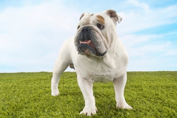
Whenever you notice that something may be wrong with your dog, it can be quite alarming.
Even just thinking about all of the various illnesses and injuries that your English Bulldog may one day face is enough to keep you up at nights and fill you with dread.
While it’s perfectly normal to worry about your Bulldog, take comfort in knowing that a lot of what you worry about may never come to pass.
In fact, a lot of conditions, like cherry eye, can be managed, treated, and even often cured.
What is cherry eye in English Bulldogs? Cherry eye is when the lacrimal gland of the nictitating membrane slips out of its normal position and visibly protrudes. Common in Bulldogs, it appears as a red or pink, fleshy mass in the eye’s inner corner and is thought to be an inherited condition.
Understanding more about the problem of cherry eye will help to relieve your fears so that you can get your dog the proper help he needs. Let’s take a closer look at this common Bulldog condition.
What Is Cherry Eye?
A veterinarian would describe cherry eye as the prolapsed gland of the third eyelid. Before we dive into the problem of cherry eye, we should first discuss the third eyelid.
Also called the nictitating membrane, the third eyelid is a translucent membrane that is drawn across the eye horizontally to moisten and protect the eye without impairing vision as regular blinking does.
In addition, the third eyelid contains one of the two lacrimal (tear) glands of the eye and is responsible for an estimated 30– 50% of the eye’s tear production.
It is also thought to provide some level of immune protection for the eye due to the presence of lymphoid tissue.
Fun Fact: Nictitating membranes are found in many mammals including dogs and cats as well as in birds, reptiles, amphibians, and fish.
Under normal conditions, you probably never notice your dog’s third eyelid or only occasionally catch a glimpse of it, and this is how it should be.
However, the third eyelid can develop issues, such as cherry eye, which make it prominent in appearance and uncomfortable for your Bulldog.
English Bulldogs experience cherry eye when the lacrimal gland pops out of its proper position and appears as a fleshy, protruding mass in the inner corner of the eye.
Because the gland is no longer where it belongs, swelling may occur due to improper blood circulation, and the normal rate of lubricating tear production may be affected.
Cherry eye is most commonly seen in dogs less than two years old and in brachycephalic breeds such as Pugs, French Bulldogs, Boston Terriers, Cocker Spaniels, and, yes, English Bulldogs.
Toy and teacup varieties of dogs also run a high risk of developing cherry eye, but this condition can affect any dog, whether purebred or of mixed lineage, at any age.
Possible Causes
In many cases, the cause of cherry eye is thought to be genetic with the tendency for suffering from this condition handed down from generation to generation.
For this reason, English Bulldogs who have experienced cherry eye at least once in their life should not be bred.
While the exact cause of cherry eye is not thoroughly understood, veterinarians agree that weak connective tissues within the eye are to blame.
Many of the brachycephalic breeds are genetically predisposed to abnormally weak connective tissue to hold the lacrimal gland securely in place, hence the high occurrence of cherry eye in short-nosed breeds.
When tissues weaken to the point of no longer being able to keep the tear gland firmly in its rightful place, the gland shifts in position and “pops” out, resulting in a classic presentation of cherry eye.
Be aware that once cherry eye has occurred, the probability rises that the other eye will experience it as well.
How to Identify
Cherry eye is easily identified by a red or pink, bulging, fleshy mass which suddenly appears in one or both inner corners of the eyes.
There is no need to panic if your Bulldog does develop cherry eye, as it is usually not an emergency situation. It just looks a whole lot worse than it actually is.
Remember that dogs are skilled at picking up on our emotions, so remaining calm is one of the best things you can do for your dog.
Cherry Eye Treatment
The Tucking Method
The most common treatment is known as the tucking method which involves stitching the errant gland back into position.
This is the least invasive procedure but may need to be repeated should the stitch become loose or fail entirely.
The Pocket Technique
The other, newer option is called the pocket technique or imbrication. For this procedure, a wedge of tissue from the area above the gland is removed thus creating a pocket to hold the gland, and the area is secured with stitches.
Both surgeries have a better chance of success if performed soon after the prolapse before too much swelling has occurred.
Can At-Home Treatments Help?
While spontaneous recovery is rare, cherry eye has been known to resolve itself, though many Bulldog owners have successfully helped the lacrimal gland slip back into position with a simple massage.
To be effective, this treatment must be done soon after the condition is noted.
The massage method involves placing repetitive, gentle pressure in a downward, diagonal direction with a damp compress to encourage the gland to move back into place.
Can Cherry Eye Be Prevented?
Sadly, there is no way to prevent cherry eye from occurring. Your best bet to avoid the issue is to purchase a Bulldog from a well reputed breeder who refuses to breed any Bulldog who has suffered from this condition.
Other Common Eye Conditions in English Bulldogs
Unfortunately, cherry eye is not the only eye condition that English Bulldogs endure. The structure of the eye is quite complex, yet delicate, presenting the opportunity for many conditions to arise.
Let’s take a quick look at the more common eye problems found in Bulldogs.
Eyelid and Eyelash Abnormalities
Though these conditions are similar in that they all involve the eyelid and lashes, they are not identical and will require different treatment.
- Entropion is defined by the eyelid turning in towards the eye thus causing the eyelashes to painfully rub against the eye itself.
- Ectropion occurs when the lower eyelid sags down, exposing delicate tissue and resulting in dryness, inflammation, and discharge.
- Distichiasis is when eyelashes grow in abnormal places (often from the meibomian glands) on the eyelid and irritate the eye.
- Ectopic cilia involves the growth of eyelashes through the inside of the eyelid directly towards the cornea.
- Trichiasis is a condition in which one or several eyelashes grow towards the eye instead of turning outwardly.
All of these conditions can cause profuse tearing, rapid blinking, twitching, squinting, and irritation which the dog may try to alleviate by pawing or rubbing the eye.
Left untreated, these disorders may lead to inflammation, corneal ulcers and/or other damage, infection, and even vision loss.
Infection and Allergies
Bacterial and viral infections, such as conjunctivitis, as well as allergic responses can cause your dog’s eyes to become red, watery, and irritated. Puffy eyelids, inflammation, and gooey discharge may be present also.
Brachycephalic Ocular Disease
This is a syndrome encompassing various ocular abnormalities and problems which stem from the unique anatomy of brachycephalic breeds.
Corneal Ulcers
Blunt trauma, scratches or cuts, irritating substances, infections, and disease can all cause a corneal ulcer.
Loosely defined, a corneal ulcer is an open sore on the cornea often accompanied by inflammation. Severity and symptoms can range from mild to severe with treatment dependent on the depth of the injury.
Dry Eye Syndrome
English Bulldogs are one of the breeds that are particularly prone to developing dry eye syndrome, or keratoconjunctivitis sicca (KCS).
KCS is caused by a lack of adequate tear production to protect the surface of the eye and the lining of the lids.
Symptoms of KCS include rapid blinking, swelling of the eye tissues and blood vessels, protruding third eyelid, itching, irritation, and mucus accumulation.
")
")
")
")